
中

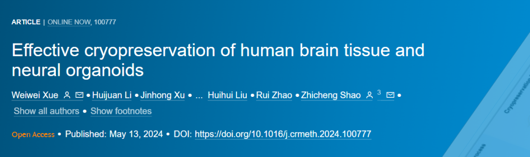
Shao Zhicheng's team from Fudan University successfully resurrected human brain organs that had been frozen for 18 months, breaking the record in the field of cryogenic technology and appearing in the Cell journal.
Cryopreservation is an effective method to preserve biological samples at low temperature, so that they can maintain their structure and function during long-term storage. In recent years, cryopreservation technology has been widely used in the field of neuroscience, especially for the preservation of human brain tissue and nerve organs.
Human brain tissue and nervous organs are valuable resources for the study of nervous system development and diseases. However, these tissues are very fragile and easily degraded at room temperature. Cryopreservation can effectively prevent the degradation of these tissues and enable them to be studied in the future.
This paper will introduce the effective cryopreservation methods of human brain tissue and nerve organs. The basic principles and steps of cryopreservation are described. Then, the factors affecting the effect of cryopreservation were discussed, including freezing rate, use of protectant and thawing process. Finally, some latest techniques for cryopreservation of human brain tissues and nervous organs are introduced.
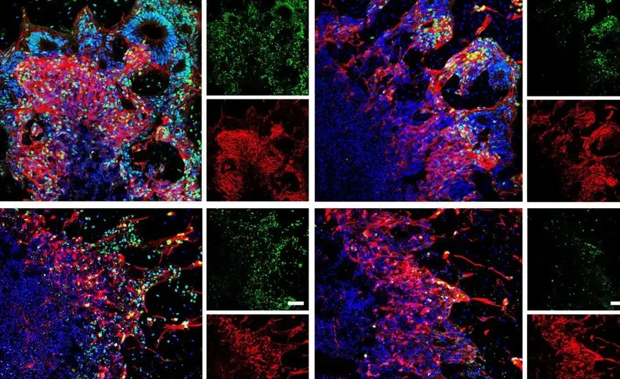
Experimental procedure.
This technology is a major breakthrough in the field of cryogenic technology and paves the way for improving the research methods of nervous system diseases. This work by Dr. Shao Zhicheng of Fudan University was also officially published in the Cell sub-journal this month.
Human brain tissue models and organ-like organs are very important in studying and simulating human nervous system diseases. However, the high maintenance cost of long-term cultured organs limits their wide application. Therefore, it is urgent to develop cryopreservation methods for brain tissue and organ-like organs.
In this study, we established a method of cryopreservation of cortical organs using methylcellulose, ethylene glycol, DMSO and Y27632 (called MEDY). This method does not destroy the structural or functional activity of nerve cells.
MEDY can be used in a variety of brain region-specific organs, including dorsal / ventral forebrain, spinal cord, optic vesicle brain and brain organs derived from patients with epilepsy. In addition, MEDY can also realize the cryopreservation of human brain tissue samples and retain the pathological features after thawing. Transcriptome analysis showed that MEDY could protect synaptic function and inhibit endoplasmic reticulum-mediated apoptosis. MEDY will achieve large-scale and reliable storage of various neurological organs and living brain tissues, and promote extensive research, medical applications and drug screening.
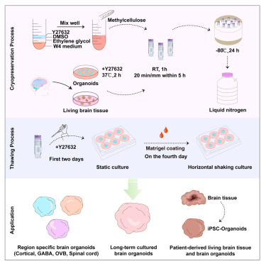
Immunofluorescence staining imaging technique shows thawed brain organs.
In response, Professor Jo ã o Pedro Magalh ã es of the University of Birmingham said he was shocked.
You know, brain cells are very fragile and extremely sensitive to stress. It is a miracle that the method adopted by the team can successfully prevent cell death and even allow them to retain their function.
Professor Magalh ã es boldly predicts that we can now imagine a scenario where, decades or centuries later, terminally ill patients can be frozen and wait for the day when there is a cure. Astronauts can be frozen and wake up to be sent to other galaxies.
Why can MEDY preserve fragile brain cells?
MEDY can effectively preserve fragile brain cells, mainly because it uses specific combinations of compounds that protect cells from damage during freezing. Specifically, MEDY includes methylcellulose, ethylene glycol, DMSO and Y27632, which work together to:
Provide physical protection: methylcellulose can form a protective layer to reduce the physical damage of ice crystals to cells.
Maintain osmotic balance: ethylene glycol and DMSO are used as penetrating cryoprotectants to help cells maintain osmotic balance and prevent cells from dying due to dehydration. Inhibition of cell death: as an inhibitor of ROCK, Y27632 can inhibit the apoptosis pathway induced by freezing and improve the cell survival rate.
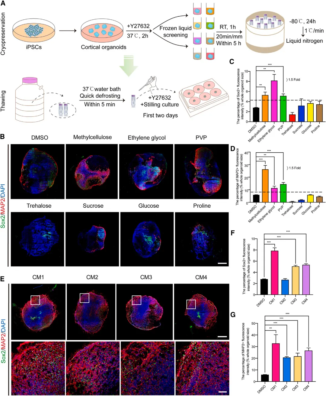
Figure 1. Establishment of MEDY cryopreservation technology
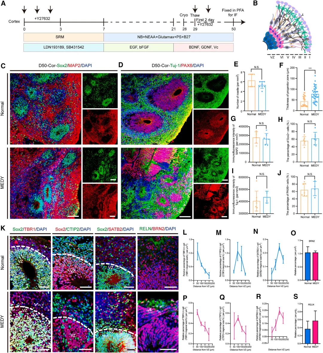
Figure 2. Protective effect of MEDY on the functional structure of cortical organs
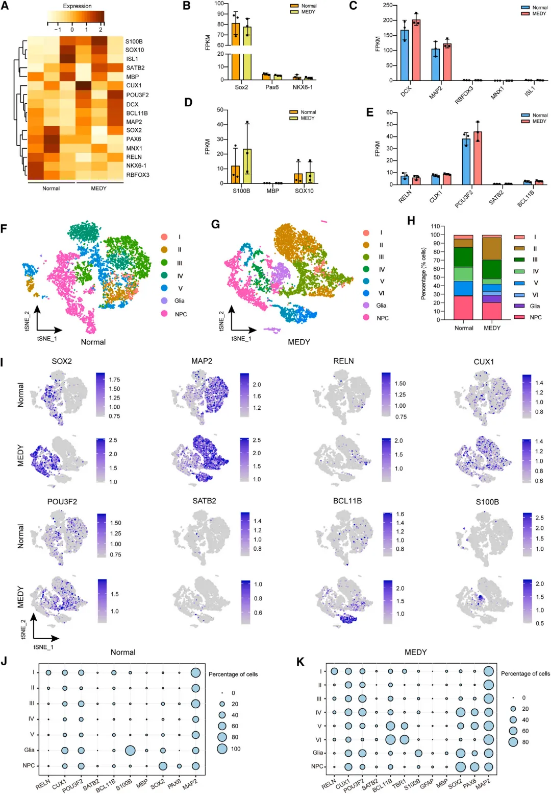
Figure 3. RNA sequencing and single cell sequencing analysis of cell diversity in normal and MEDY cortical organs
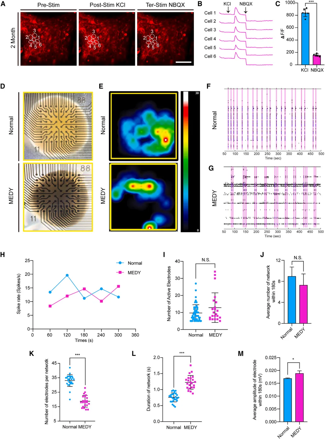
Figure 4. Detection of functional activity of normal and MEDY cortical organs by calcium imaging and MEA
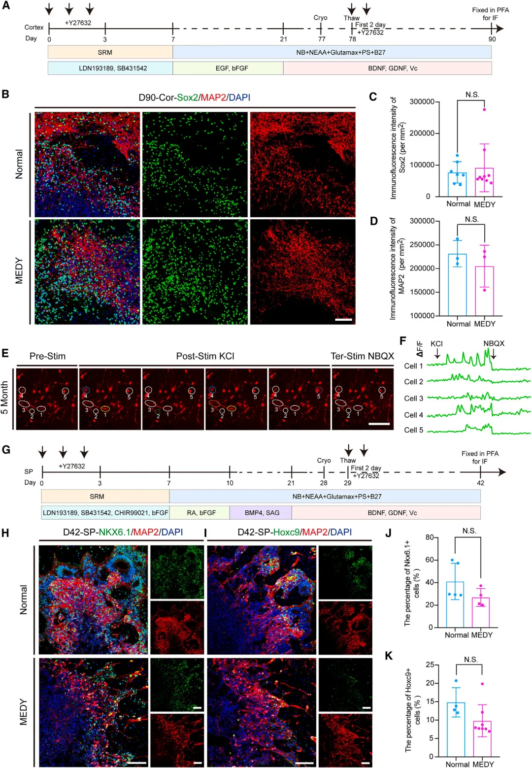
Figure 5. MEDY cryopreservation can be used for the protection of long-term cultured cortical and SP organs
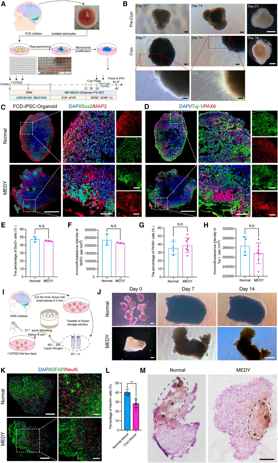
图 6. MEDY 冷冻保存适用于癫痫儿童来源的皮质类器官和活脑组织的保存
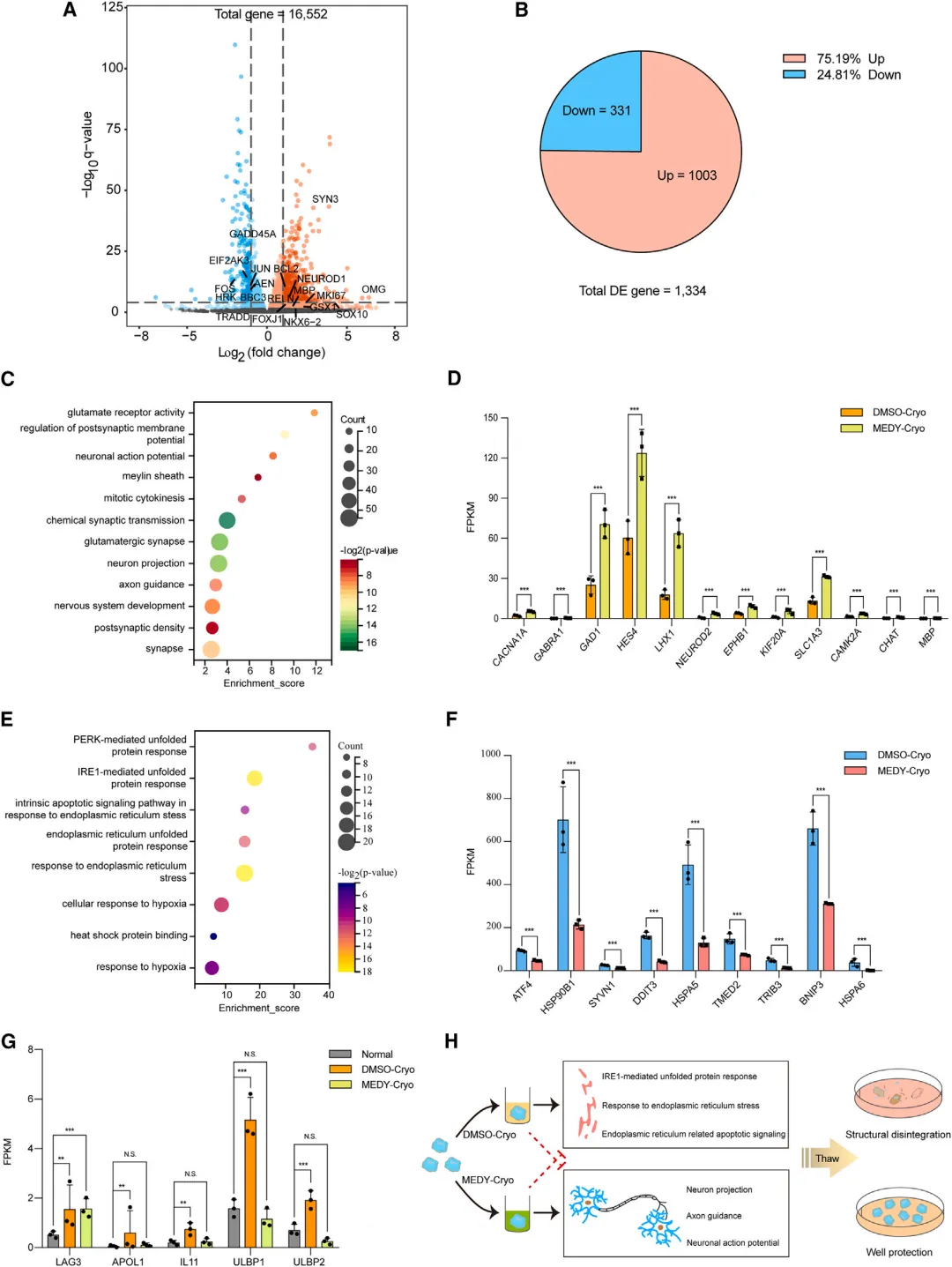
Figure 7. RNA sequencing reveals changes in gene expression behind MEDY cryopreservation
Article source: https://doi.org/10.1016/j.crmeth.2024.100777