
中

The wave of regenerative medicine is surging forward. Induced pluripotent stem cell therapy, with its immense application potential and a continuous stream of breakthrough achievements, is rapidly becoming the hottest field in the global medical sector.
Recently, exciting progress has been made in this cutting-edge field - not only has it opened up new paths for the treatment of intractable diseases, but it has also injected strong impetus into the development of regenerative medicine as a whole, making the hope of overcoming difficult diseases increasingly clear.
Cell Stem Cell | Generation of iPSCs for Use in Tissue Engineering Vascular Catheters (TEVCs)
Recently, the research team from the Cardiovascular Research Center of Yale University was published in the top journal "Cell Stem Cell", proposing a brand-new construction plan for tissue-engineered vascular conduits (TEVCs) - using decellularized human umbilical arteries as the natural scaffold, combined with endothelial cells derived from human induced pluripotent stem cells (hiPSCs), to create a fully biologic vascular conduit with immediate antithrombotic function and rapid host integration. This brings revolutionary treatment hope to patients with congenital heart diseases and other vascular disorders.
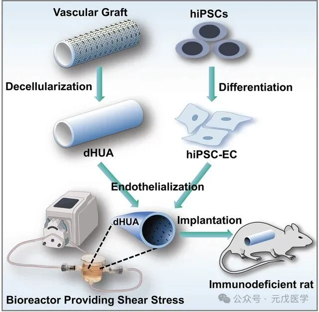
Graphical Summary
For children with single ventricle congenital heart disease, the vascular catheter is the "lifeline" that sustains their lives. However, for decades, traditional synthetic grafts have been plagued by three major problems: thrombosis, frequent infections, and the inability to grow with the body. As a result, countless children have had to undergo repeated surgeries to replace the catheter. Therefore, developing a new type of vascular graft that possesses immediate antithrombotic properties, host integration capabilities, and low immunogenicity has become the core scientific issue in this field.
The core breakthrough of this research lies in completely discarding synthetic scaffolds and inflammatory-prone bone marrow cells, and constructing a new generation of vascular conduits that are entirely based on biomaterials and stem cells:
01 Natural Umbilical Artery Stent: Say Goodbye to the Inflammatory Nightmare of Synthetic Materials
The research team did not use traditional synthetic materials such as polytetrafluoroethylene or polyester. Instead, they used decellularized human umbilical arteries (dHUAs) as scaffolds. Through a special process, all the cells in the umbilical arteries were removed, leaving only the natural extracellular matrix (ECM) framework. This not only completely retained the original three-dimensional structure and excellent mechanical properties of the blood vessels, but also eliminated the chronic inflammatory response caused by synthetic materials at its root.
The study selected dHUA as the natural scaffold, and its advantages lie in:
02 hiPSC-derived endothelial cells: An "off-the-shelf" supply of "customized cells"
Human induced pluripotent stem cells (hiPSCs) can proliferate indefinitely and differentiate into various cell types of the human body. The research team directed the differentiation of hiPSCs into endothelial cells (hiPSC-ECs), providing a consistent and scalable cell source for vascular conduits.
The study used hiPSC-ECs instead of the previous bone marrow-derived cells. The scientific basis for this is as follows:
Source standardization: hiPSC lines can establish master cell banks (MCB) and working cell banks (WCB) under GMP conditions, achieving consistency between batches.
Differentiation accessibility: Through the targeted induction by factors such as VEGF-A/FGF-2/BMP-4, a high-purity population of CD31⁺/VE-Cadherin⁺ endothelial cells can be obtained.
Functional controllability: Prevents the excessive proliferation phenotype of bone marrow EPCs, reducing the risk of lumen stenosis.
Utilizing human induced pluripotent stem cells (hiPSCs) to develop tissue-engineered vascular conduits (TEVCs), for the implantation of vascular grafts as replacements for the inferior vena cava in nude mice
03 Gradient shear force training: Making endothelial cells "ready to work earlier"
In order to enable hiPSC-ECs to better adapt to the blood flow environment within the body, the research team subjected the cells to gradient shear force training in a bioreactor.
Training Plan:
Bioreactor simulates arterial blood flow conditions
The shear stress gradient increased to 15 dynes/cm² (the level of a normal artery)
The pre-transplantation adaptive setting was reduced to 5 dynes/cm² (matching the implantation site of the inferior vena cava)
Molecular mechanism basis: Shear force activates downstream signaling pathways through mechanosensors on the endothelial cell surface (such as the Piezo1 ion channel, VE-cadherin/PECAM-1 complex), and upregulates:
Expression of nitric oxide synthase (eNOS) → Promotes vasodilation
Expression of thrombomodulin → Enhancement of anticoagulant function
Expression of tissue-type plasminogen activator (t-PA) → Promotes fibrinolysis
04 Instant antithrombotic function: No need for lifelong anticoagulation
This is one of the most clinically valuable breakthroughs of this research. In previous cases of vascular catheter transplantation using tissue engineering, it was necessary to wait for the host endothelial cells to cover the vessel to exert the anti-thrombotic effect, and during this period, anticoagulant drugs had to be relied upon. However, the endothelialized TEVCs constructed by the Yale team could immediately exert anti-thrombotic protection after transplantation, significantly reducing the risk of bleeding related to anticoagulants.
15–5 dynamic/centimeter² progressive shear flow training can enhance the function of hiPSC-endothelial cell-derived vascular endothelium in the implanted inferior vena cava (IVC) grafts.
05 Rapid Host Integration: Achieving True "Vascular Regeneration"
The transplanted TEVCs do not persist as "foreign substances" permanently; instead, they can quickly recruit the host's own endothelial cells and vascular smooth muscle cells.
The research team conducted a comprehensive test of this new type of TEVCs in a rat inferior vena cava replacement model, and the results were very encouraging:
No thrombosis formation: After transplantation, all TEVCs remained unobstructed and no thrombosis was observed. In contrast, all the grafts in the control group that did not undergo shear force training developed thrombosis and became blocked.
Complete re-endothelialization: One month after the surgery, the surface of the graft was completely covered by host endothelial cells, and the expression levels of endothelial markers CD31 and eNOS were comparable to those of natural blood vessels.
Extremely low inflammatory response: Two months after the surgery, there were almost no macrophages detected in the graft, indicating that the inflammatory response has been significantly suppressed.
The method is highly versatile: TEVCs constructed using different hiPSC cell lines all demonstrated consistent antithrombotic function and host integration ability, proving that this technology has broad applicability.
Although this research has achieved significant breakthroughs, there is still a long way to go before it can be applied in real clinical settings. Several key issues still need to be addressed:
Verification through large animal and human trials: Currently, the research has only been conducted in small animal models. Long-term durability and safety need to be verified in large animal models, and human clinical trials will be gradually carried out.
The mechanism research needs to be further explored: The specific molecular mechanisms by which hiPSC-ECs regulate antithrombotic function and integrate with host cells have not yet been fully elucidated.
Comprehensive assessment of mechanical properties: A more systematic analysis is needed for the long-term mechanical stability of the graft and the vascular reactivity function, etc.
Cell type-specific optimization: Whether the artery and vein-specific hiPSC-ECs will affect the performance of the grafts still requires further research.
Summary
This study successfully constructed a fully biologic TEVCs with immediate antithrombotic function and host integration ability by integrating three key technical elements: natural ECM scaffolds, hiPSC-derived endothelial cells, and biomechanical function training. This strategy effectively overcame the limitations of previous synthetic scaffolds and bone marrow-derived cells, providing a promising tissue engineering solution for clinical treatment of SVCHDs and other vascular reconstruction indications.