
中

Source: BioArt
As the only means to achieve complete totipotency of somatic cells so far, somatic cell nuclear transfer has broad application value in the fields of biology and regenerative medicine. In the past few decades, this technique has been successful in more than 20 mammal species, including primates, but there are a variety of epigenetic obstacles in the reprogramming of somatic reconstructed embryos, resulting in a very low birth rate of cloned animals and accompanied by developmental defects of the fetus and placenta.[1]。
In recent years, many researchers have tried to improve the success rate of nuclear transfer by correcting the abnormal apparent modification in cloned embryos. By studying the mechanism of reprogramming of nuclear transfer embryos, Gao Shaorong of Tongji University and Zhang Yi of Harvard Medical School have identified the barriers to reprogramming caused by abnormalities of histone modifications (H3K9me3, H3K4me3 and H3K9ac) during preimplantation embryo development. Effective intervention can greatly enhance the preimplantation developmental potential of nuclear transfer embryos [2-5].
In addition, abnormal activation of Xist and abnormal expression of H3K27me3-dependent imprinted genes in nuclear transfer embryos can lead to defects in embryonic development after implantation. [6,7] Restoring their normal expression level by gene editing technique can improve the developmental ability and birth rate of cloned embryos after implantation.[6,8]
However, on the basis of greatly improving the blastocyst rate and post-implantation development ability of nuclear transfer technology, the final birth rate is still much lower than that of normal or IVF embryos.This means that there are many other unknown obstacles that limit the ability to clone embryo development. Looking back to the whole development process of nuclear transfer technology, most studies mainly focus on the abnormal development of cloned embryos before and after implantation. Due to the lack of research means and tools, little is known about the developmental regulation mechanism of the peri-implantation stage of cloned embryos.
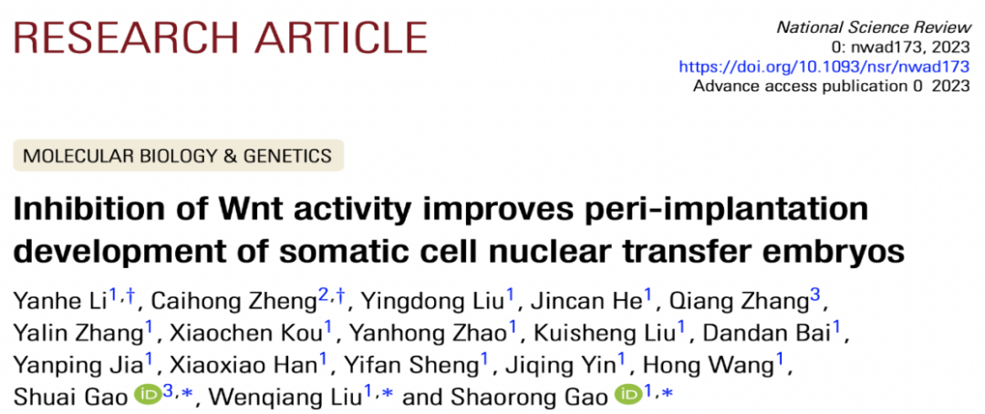
On August 16, 2023, Gao Shaorong, School of Life Science and Technology, Tongji University and Gao Shuai's research group of China Agricultural University published a research paper entitled Inhibition of Wnt activity improves peri-implantation development of somatic cell nuclear transfer embryos on National Science Review online. This study identified a new epigenetic disorder that leads to the failure of peri-implantation cloned embryos: abnormal activation of the Wnt pathway. More importantly, intervention and inhibition of Wnt signaling pathway (Wnti) by adding inhibitors of Wnt pathway to the culture medium can significantly improve the developmental ability of cloned embryos during peri-implantation period and significantly increase the birth rate of cloned mice.
It is worth mentioning that in another work published at the same time, Gao Shaorong's team explained that abnormal histone modification affected the pedigree establishment of early cloned embryos, suggesting the importance of the study of peri-implantation development of cloned embryos after the first pedigree differentiation. However, the inaccessibility of the growth of peri-implantation embryos in the maternal uterus has seriously restricted the study on the regulation mechanism of the development of cloned embryos during peri-implantation.
So how are the morphogenesis, signal transduction and apparent regulation events of cloned embryos carried out at this stage? In order to answer this question, Gao Shaorong's research group optimized and established a 3D embryo culture system for in vitro culture of cloned embryos, which can simulate the development process of peri-implantation in vivo and realize the visualization of peri-implantation development of cloned embryos.
Combined with in vitro culture and in vivo transplantation experiments, it was found that most of the cloned blastocysts showed developmental arrest at the E4.5-E5.25 stage, which was characterized by obvious abnormalities in the transformation from inner cell mass (inner cell mass,ICM) to ectoderm (epiblast, EPI) oval barrel structure (egg cyclinder).
The morphological transformation of ectoderm and the implantation rate of embryos in vivo could be greatly corrected by supplementing cloned blastocysts with ICM cells of embryos fertilized in vitro, while ICM cells supplemented with cloned embryos had no effect on the development of peri-implantation embryos.
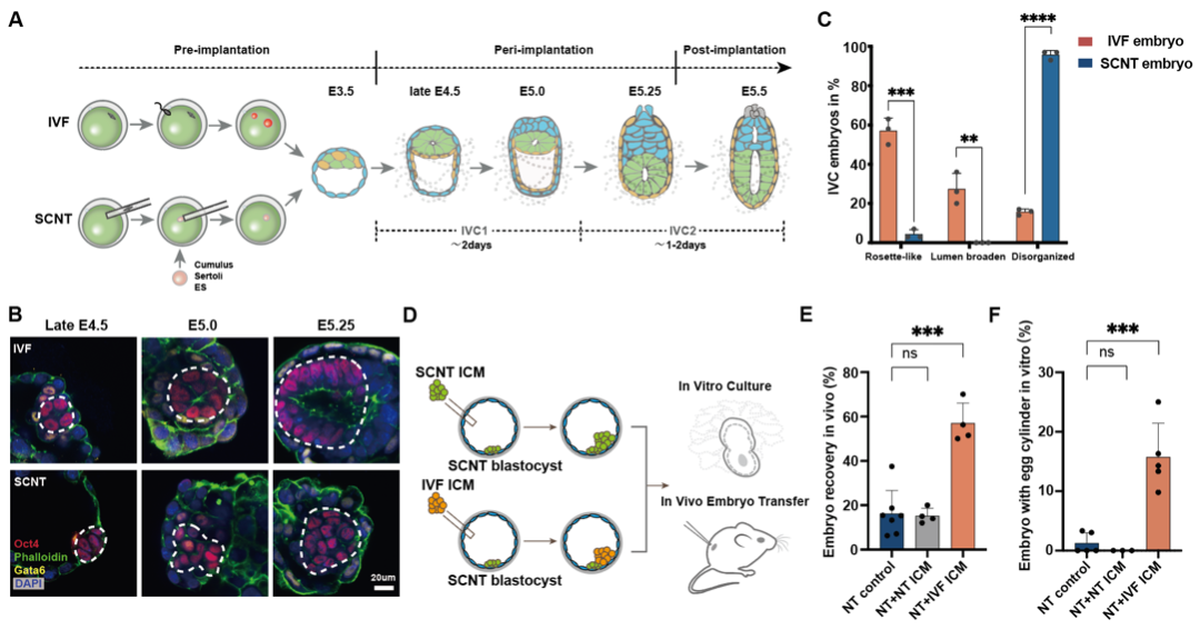 Figure 1: abnormal morphological process of ectoderm in cloned embryos during peri-implantation
In order to verify the molecular mechanism of abnormal development of EPI, the researchers conducted a large number of / single cell transcriptome analysis of embryos cultured in vitro and in vivo during peri-implantation. The results showed that the transition of Na ï ve-Primed of cloned embryos was blocked compared with that of embryos fertilized in vitro, and this abnormal phenomenon was common in different donor cells (granulosa cells, Sertoli cells). The peri-implantation development of cloned embryos derived from embryonic stem cells. Further analysis of trace histone ChIP-seq data showed that the H3K27me3 remodeling of the promoter region of differentially expressed pluripotent genes was also abnormal.
In order to find out the causes of abnormal pluripotency transition, the researchers conducted comparative transcriptional group, reporter system and signal pathway intervention experiments, and confirmed that the persistent abnormal activation of Wnt pathway is an important obstacle to the pluripotent state transition of cloned embryonic EPI cells.
The researchers further found that many Wnt pathway antagonists, such as Dkk1, Tcf7l1 and other genes, were significantly inhibited in the preimplantation blastocyst stage.
Through the analysis of public data of histone modification in mouse donor cells and early embryos, it was found that H3K27me3 carried in donor cells may be the root cause of antagonistic gene expression inhibition of Wnt pathway such as Dkk1 during implantation.
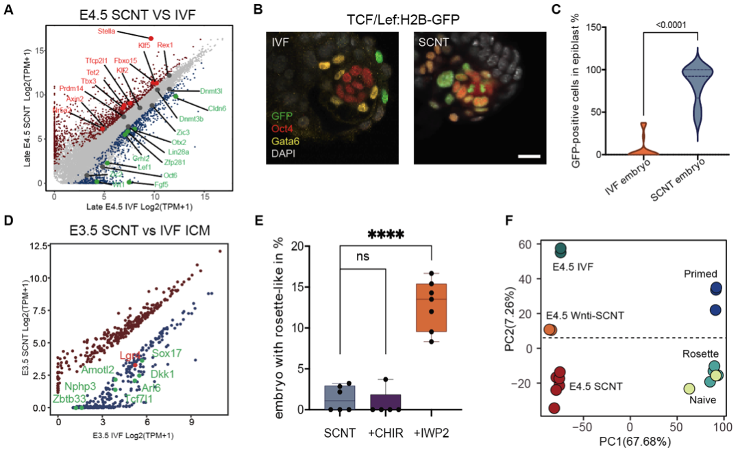 Figure 2: abnormal activation and intervention of Wnt pathway in ectoderm of cloned embryos during peri-implantation
Then, in order to verify the improvement of embryo development by inhibiting the continuous activation of Wnt pathway during peri-implantation, IWP2/IWR1-endo, an inhibitor of Wnt pathway, was added to the culture medium to interfere with cloned blastocysts. It was found that Wnt pathway inhibitor could significantly promote the transformation of pedigree development, EPI morphogenesis, apparent state and pluripotent state of cloned embryos during peri-implantation. Importantly, cloned blastocysts treated with Wnt pathway inhibitors can significantly improve embryo implantation ability and birth rate (granulosa cell donor nuclear transfer embryo birth rate increased from 0.72% to 5.55-7.04%; supporting cell donor nuclear transfer embryo birth rate increased from 0% to 10.3%).
Finally, through the combination of important apparent repair methods reported in the past, the researchers found that the combination of Xist KO and Dnmt3a/b interference could further increase the birth rate of nuclear transfer blastocysts to more than 20%.
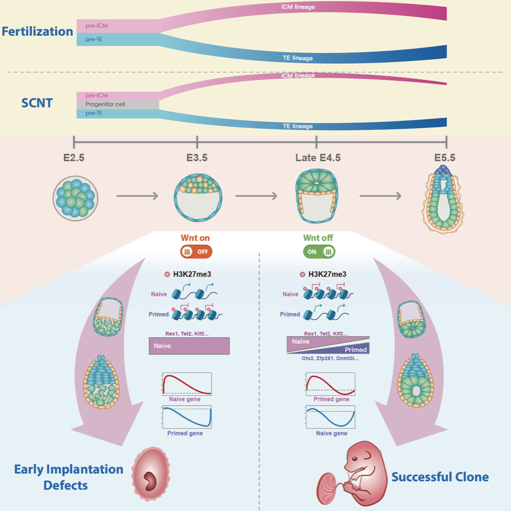 Figure 3: obstacle mechanism of abnormal development of cloned embryos during peri-implantation
In general, this study uncovered the dynamic development process of cloned embryos during peri-implantation period for the first time by using the optimized blastocyst simulated implantation culture system, and revealed the abnormal biological events of ectoderm morphogenesis and pluripotency transformation during this period. Importantly, the researchers found that the continuous activation of Wnt pathway is an important obstacle to the peri-implantation development of cloned embryos. The intervention of Wnt activation by small molecular inhibitors and other apparent repair methods can further improve the efficiency of nuclear transfer. This study provides an important theoretical basis for understanding peri-implantation embryonic development and expanding the application of cloning technology in developmental biology, regenerative medicine and animal husbandry.
Dr. Li Yanhe of Tongji University and Zheng Caihong, associate researcher of Beijing Genome Research Institute, are the co-lead authors of this study. Professor Gao Shaorong and Professor Liu Wenqiang of Tongji University and Professor Gao Shuai of China Agricultural University are the co-authors.
Original link: https://doi.org/10.1093/nsr/nwad173
References:
1. Matoba, S. and Y. Zhang, Somatic Cell Nuclear Transfer Reprogramming: Mechanisms and Applications. Cell Stem Cell, 2018.
2. Liu, W., et al., Identification of key factors conquering developmental arrest of somatic cell cloned embryos by combining embryo biopsy and single-cell sequencing. Cell Discov, 2016. 2: p. 16010.
3. Matoba, S., et al., Embryonic development following somatic cell nuclear transfer impeded by persisting histone methylation. Cell, 2014. 159(4): p. 884-95.
4. Yang, G., et al., Dux-Mediated Corrections of Aberrant H3K9ac during 2-Cell Genome Activation Optimize Efficiency of Somatic Cell Nuclear Transfer. Cell Stem Cell, 2020.
5. Gao, R., et al., Inhibition of Aberrant DNA Re-methylation Improves Post-implantation Development of Somatic Cell Nuclear Transfer Embryos. Cell Stem Cell, 2018. 23(3): p. 426-435 e5.
6. Inoue, K., et al., Impeding Xist expression from the active X chromosome improves mouse somatic cell nuclear transfer. Science, 2010. 330(6003): p. 496-9.
7. Matoba, S., et al., Loss of H3K27me3 Imprinting in Somatic Cell Nuclear Transfer Embryos Disrupts Post-Implantation Development. Cell Stem Cell, 2018. 23(3): p. 343-354 e5.
8. Wang, L.Y., et al., Overcoming Intrinsic H3K27me3 Imprinting Barriers Improves Post-implantation Development after Somatic Cell Nuclear Transfer. Cell Stem Cell, 2020. 27(2): p. 315-325 e5.