Source: BioArt
The normal development of human embryos after implantation largely depends on the morphogenesis of the outer tissue of the embryo and its influence on the cell tissue of the embryo. However, due to ethical and technical challenges, there is still a lack of models that can reflect the morphogenesis of the spatial tissue of human embryos after implantation, and the construction of such models is crucial for solving problems such as fertility and embryonic development defects.
On August 1, 2022, Jacob H. Hanna's team at the Weizmann Institute in Israel published a study in the journal Cell, demonstrating that mouse primitive embryonic stem cells (naive ESCs) have the ability to self-assemble into embryos with spatial tissue morph outside the uterus (previously known as sEmbryos). It was later renamed SEMs [1]. Inspired by this, they wanted to test whether they could induce human pluripotent stem cells to develop dynamically extrauterine to the gastrula stage.
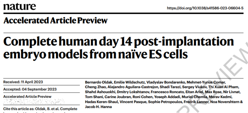
On September 6th, 2023, Jacob H. Hanna's team published an article in the journal Nature entitled "Complete human day 14 post-implantation embryo models from naive ES cells." To demonstrate that unmodified human naive ESCs can also be induced to self-assemble into SEMs that reproduce almost all known lineages and structures of postimplantation human embryos (e.g. upper and lower derm, etc.) and dynamically simulate key developmental features from 7-8 days to 13-14 days after fertilization. The construction of this platform will help people understand the unknown events in the early stages of human embryo implantation.

Similarly, in mice, if you want to obtain Primitive endoderm (PrE) and Trophoblast stem cells (TSC) lineages from naive PSCs, Rapid induction by transient overexpression of transcription factors Gata4 or Cdx2 is required under optimal conditions. Human embryos (but not mouse embryos) will form ExEM structures after implantation, so the authors induced human ESCs with human PrE and ExEM regulatory factors GATA4 and GATA6, respectively, and used FACS staining markers (PDGFRa+) to determine the optimal induction conditions. At the same time, human ESCs were induced into trophoblast (Tb) with CDX2, and the induced TB-like cells and PrE/ EXEM-like cells were assembled into a complete embryoid structure by calibrating the aggregation conditions of these cells, such as the number, proportion and medium composition required at different stages.
 Fig. 1. Complete self-assembly of human naive ESCs into SEM and SEM structure bright field diagram under in vitro culture conditions.
Cultured outside the uterus for 3-4 days, these cells spontaneously formed 3D spherical structures and reflected the structural characteristics of TB-like cells wrapped in the outermost part of the SEM when implanted in the uterus, and the EPI-like and HYP-like cells expressing OCT4 and SOX17, respectively, began to form amniotic cavity (AC) in the 6th mesodermal compartment. The hypodermoid compartment begins to form a yolk sac-like cavity (YS), and a double-layer disk-like structure is formed between the two, similar to the embryonic state of 9-10 dpf in humans. On this basis, they described in more detail the developmental characteristics of key lineages in human SEMs, such as immunostaining of TFAP2A and ISL1 in SEM to confirm the amniotic features of EPI-like chambers in terms of location, morphology, and gene expression. In addition, the researchers also used Chromium 10X scRNA-seq to perform single-cell transcriptomic analysis of SEMs, and based on the expression of cell-specific markers to confirm the validity of each cell identity definition in the SEM. Transcription profiles were compared with six human embryo datasets [2-7] to verify the similarity of SEMs to early human embryo cell types.
In short, this work produces a human SEMs outside the uterus that can self-organize and reproduce the 3D structure and key developmental markers of the early embryo in utero, without providing directional signal induction in the process. Although the model is not exactly the same as the human embryo, it also helps to realize the purpose of studying the key events of embryonic development. In this study, the team observed the problems of "low efficiency" and "variable development stage (unstable)" in the construction of human SEM. In the future, while further optimizing the culture conditions, it is necessary to further test whether the model can continue to complete the development of gastrula stage and form various organs.
Original link:https://doi.org/10.1038/s41586-023-06604-5
Reference:
1.Tarazi, S. et al. Post-gastrulation synthetic embryos generated ex utero from mouse naive ESCs. Cell 185, 3290-3306.e25 (2022).
2.Zhao, C. et al. Reprogrammed blastoids contain amnion-like cells but not trophectoderm.bioRxiv 2021.05.07.442980 (2021) doi:10.1101/2021.05.07.442980.
3. Yan, L. et al. Single-cell RNA-Seq profiling of human preimplantation embryos and embryonic stem cells. Nat Struct Mol Biol 20, 1131–1139 (2013).
4. Yanagida, A. et al. Naive stem cell blastocyst model captures human embryo lineage segregation. Cell Stem Cell 28, 1016-1022 (2021). doi:10.1016/j.stem.2021.04.031.
5. Petropoulos, S. et al. Single-Cell RNA-Seq Reveals Lineage and X Chromosome Dynamics in Human Preimplantation Embryos. Cell 165, 1012-1026 (2016).
6. Meistermann, D. et al. Integrated pseudotime analysis of human pre-implantation embryo single-cell transcriptomes reveals the dynamics of lineage specification. Cell Stem Cell 28,1625-1640.e6 (2021).
7. Xiang, L. et al. A developmental landscape of 3D-cultured human pre-gastrulation embryos. Nature 2019 577:7791 577, 537–542 (2019).